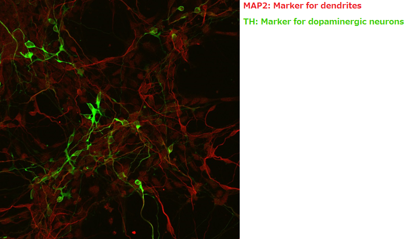
The product is a serum-free medium designed for primary culture of rat and murine neurons, and is suited for culturing cells of the central nervous system. The product contains culture supernatant of rat glial cells.


Features

Rapid production of mature neurons

Approximately 1/2 of conventional culture method

Neuron culture medium: 14 days

Conventional culture medium: approximately 1 month


Low density culture

1/5 - 1/10 the number of cells compared with conventional methods

Neuron culture medium: 0.1 ×10⁶ cells/mL

Conventional culture medium: 0.5~1.0×10⁶ cells/mL


Ready-to-Use

Cells can be cultured with the culture medium alone, with no need for additional supplements.

Confirming dendrite outgrowth

NS Basal Medium is a chemically-defined synthetic medium optimized for culturing nerve cells. NS Supplement is a serum-free supplement (containing retinyl acetate) specifically designed for supporting the growth of nerve cells. When mixed together, they can be used for culturing a variety of nerve cells, including rat hippocampal neurons and neural stem cells.

Primary Culture of Rat Hippocampal Neurons

Rat hippocampal neurons isolated from E19 embryos were cultured using poly-L-lysine-coated microtiter plates. 
The morphology of these neurons was analyzed on day 6. We also examined the expression of the neuronal markers MAP2(a+b) and the glial marker GFAP on day 21

Day 6

00578_img01.png

< Growth Medium >
NS Basal Medium containing 2% NS Supplement and 0.5 mM L-glutamine

< Seeding Density >
13,000cells/well(96well plate)

Day 21

00578_img02.gif

Primary Culture of Rat Cortical Neurons

Primary cortical neurons isolated from E17 rat embryos were cultured using poly-L-lysine-coated microtiter plates. 
The morphology of these neurons was analyzed on day 8. We also examined the expression of the neuronal markers MAP2(a+b) and the glial marker GFAP on day 14.

Day 8

00578_img03.png

< Growth Medium >
NS Basal Medium containing 2% NS Supplement and 0.5 mM L-glutamine

< Seeding Density >
60,000cells/well(96well plate)


Day 14

00578_img04.gif


00593_img01.png

The original recipe reported by the Miyawaki team in 2011 termed Scale was an aqueous solution based on urea that limited because the transparency process itself can damage the structures under study.
The research team spent 5 years improving the effectiveness of the original recipe to overcome this critical challenge, and the result is ScaleS, (we called SCALEVIEW-S) a new technique with many practical applications. SCALEVIEW-S creates transparent brains with minimal tissue damage, that can handle both florescent and immunohistochemical labeling techniques, and is even effective in older animals.
The new technique creates transparent brain samples that can be stored in SCALEVIEW-S solution for more than a year without damage. Internal structures maintain their original shape and brains are firm enough to permit the micron-thick slicing necessary for more detailed analyses.

Data provided by Dr. Hiroshi Hama, Tetsushi Hoshida and Dr. Atsushi Miyawaki, Laboratory for Cell Function Dynamics, Brain Science Institute, RIKEN Biotechnological Optics Research Team, Center for Advanced Photonics, RIKEN Cooperation with Olympus

Iba1 is a calcium-binding protein with a molecular weight of 17,000 specifically expressed in macrophage/microglia. Recently, microglia has attracted attentions because neurological damage effect by production of NO, TNF-α, and IL-1β have been proved in addition to its role in neurotrophic effects/neuroprotective actions. These products are antibodies that specifically recognize Iba1, and are available for a microglial marker.
In particular, "Anti Iba1, Rabbit (for immunocytochemistry) Code No. 019-19741" is used by many worldwide researchers as a standard for microglial marker antibody. There are also many references in major journals such as Nature, Cell etc.

Articles that you may also interested in

Search Articles

Latest Articles

  • CD112(也称为Nectin-2或脊髓灰质炎病毒受体相关2蛋白)是I型跨膜糖蛋白及Ig基因超家族成员(IGSF)。CD112...

  • Iba1是一种约17 kDa的蛋白,在神经系统的小胶质细胞上特异性表达,被用作小胶质细胞标记物。本产品是在FUJIFILM Wako的热销抗体Anti Iba1,...

  • CEPT是美国国立卫生研究院(NIH)开发的小分子混合物。由4种成分组成,并取其组成成分(Chroman 1、Emricasan、Polyamines、Trans-ISRIB)的开头字母命名为“CEPT”。 研究表明,CEPT可防止人ES/iPS细胞(人多能干细胞:hPSCs)在细胞传代中的细胞应激和DNA损伤1)。与强效ROCK抑制剂Y-27632相似,可提高细胞传代和冻存时hPSCs的存活率。 此外,与基于hPSCs的胚状体和类器官形成、单细胞克隆和基因编辑等干细胞研究中所用的现有培养方法相比,CEPT还可改善细胞存活率1,2,3)。 FUJIFILM Wako已上线经无菌测试且无需制备的即用型CEPT...